Cone beam CT (CBCT) dental scan in Casablanca
Cone beam CT, also called CBCT, dental CT scan or 3D dental X-ray, shows your teeth and bone in three dimensions. At Casa Anoual Dental Clinic, it is performed on site, on your dentist's prescription or after a consultation with Dr Zniber Mounir.
Everything about cone beam CT
What is a cone beam CT?
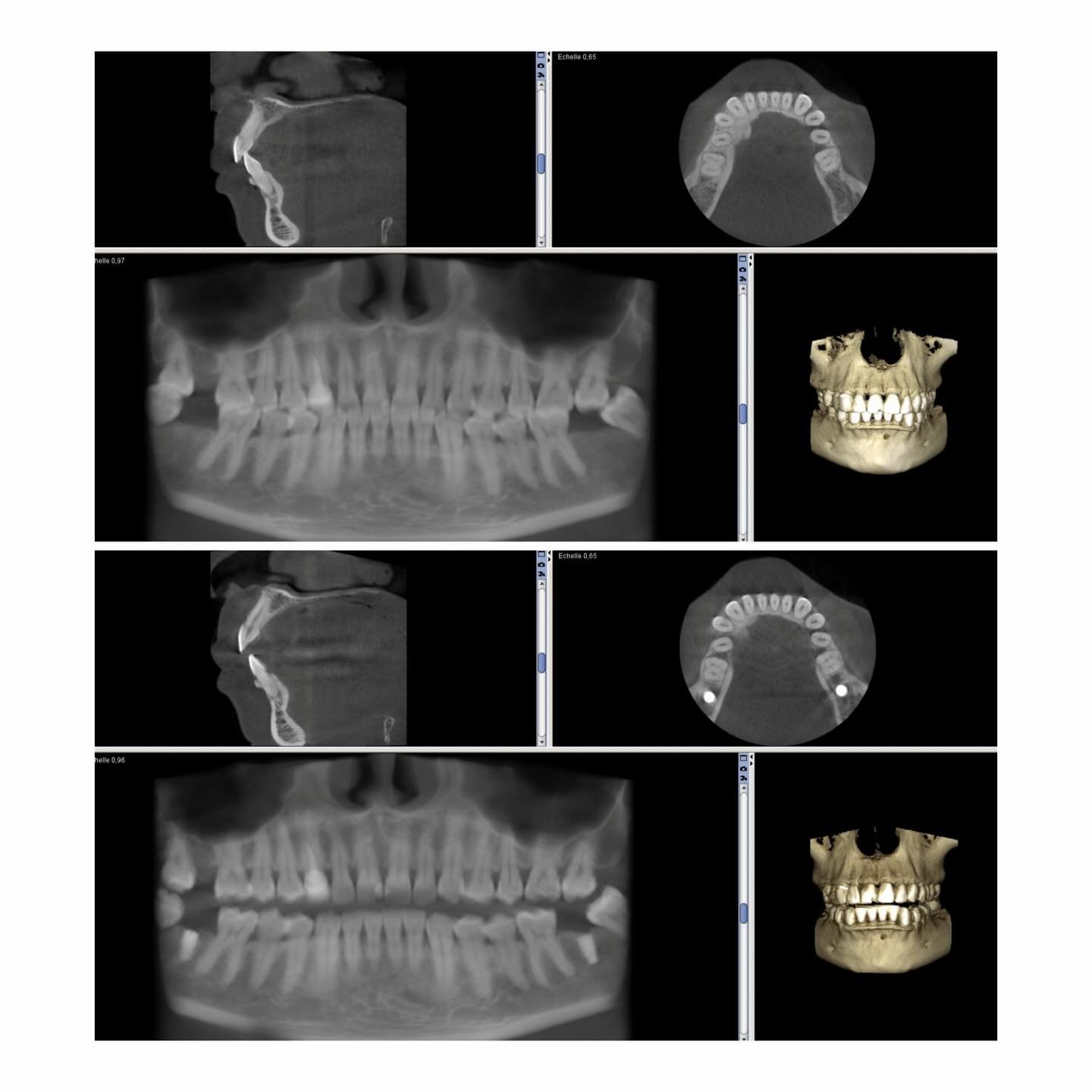
CBCT (cone beam computed tomography) is a scanner designed for the head and jaws. A cone-shaped X-ray beam rotates once around you; the computer then rebuilds a 3D volume that the dentist explores slice by slice: front, side, top, or along the dental arch.
Cone beam or medical CT?
| Cone beam (CBCT) | Medical CT (dental protocol) | |
|---|---|---|
| Device | Dedicated to head and jaws | Hospital scanner |
| Position | Seated or standing | Lying in a ring |
| Dose | Much lower | Higher |
| Detail of teeth and bone | Very fine | Good |
| Where? | At the clinic | Imaging centre |
When is a cone beam needed?
- Before an implant: bone height and width, position of the inferior alveolar nerve and sinus. Dr Zniber plans every implant on it, then designs the 3D-printed surgical guide.
- Impacted wisdom tooth or one close to the nerve, before extraction.
- Root canal treatment: extra canal, crack, lesion at the root tip, failed treatment.
- Orthodontics: impacted canine, root resorption of neighbouring teeth.
- Bone graft and sinus lift: assessment and follow-up (learn more).
- Cysts and jaw lesions, maxillary sinus, bone assessment in periodontics, checking an implant placed elsewhere.
Small, medium or large field of view
A small field covers a few teeth (root canal, one wisdom tooth); a medium field one or both arches (implants); a large field includes the sinuses or joints. The smallest useful field is always chosen: lower dose and sharper image.
The examination
- You remove jewellery, glasses and any removable appliance.
- You are positioned with your head supported, sometimes biting on a small block.
- The device rotates once around your head: stay completely still for these few seconds.
- The 3D volume is rebuilt and analysed.
The examination is painless and needs no injection.
Referred by another dentist?
The clinic also welcomes patients referred by their dentist, orthodontist or surgeon for imaging only. Simply:
- Book an appointment by phone on +212 6 97 88 85 80 or on WhatsApp, stating the prescribed examination;
- bring the prescription, an ID and, if you have them, your previous X-rays;
- leave with your images in the format of your choice: USB stick, CD, email or WhatsApp, DICOM files for your dentist's software, or printouts.
No prescription? Dr Zniber examines you first and chooses the examination you need, no more, no less.
How to prepare
- Remove jewellery, piercings, earrings, glasses and hair clips: metal creates artefacts on the image.
- Take out any removable appliance or denture, unless your dentist says otherwise.
- Tell us if you are or might be pregnant: the examination is postponed or replaced whenever possible.
- Stay still during the acquisition, which only lasts a few seconds.
Radiation dose: very low-dose examinations
Digital dental imaging uses low doses. The values below are orders of magnitude: they vary with the device, the settings and the area examined.
| Examination | Typical effective dose |
|---|---|
| Periapical (intraoral) X-ray | under 5 µSv |
| Lateral cephalometric X-ray (ceph) | under 10 µSv |
| Panoramic X-ray | about 10 to 25 µSv |
| Cone beam CT (CBCT) | about 20 to 200 µSv depending on field of view |
| For comparison: natural background radiation | about 7 µSv per day |
Each examination is only performed when it is useful for your diagnosis or treatment, with the smallest possible field of view.
Need a cone beam? Call +212 6 97 88 85 80 or send your prescription on WhatsApp.
Your questions, our answers
Cone beam, CBCT, 3D dental scan: is it the same examination?
Yes. Cone beam, CBCT, dental CT scan and 3D dental X-ray all refer to the same examination.
Is a cone beam required before an implant?
At the clinic, every implant is planned on a cone beam: it measures the bone, avoids the nerve and sinus, and is used to make the surgical guide.
How long does a cone beam take?
The rotation lasts a few seconds; allow a few minutes including positioning.
Is a cone beam dangerous?
Its dose is low, much lower than a medical CT. It is only performed when useful, with the smallest possible field.
Can I have a cone beam without being a patient of the clinic?
Yes, on your dentist's prescription. You leave with your images for your dentist.
In what format are the images provided?
In the format of your choice: USB stick, CD, email or WhatsApp, DICOM files for your dentist's software, or printouts.
Rated 4.9 stars by our patients
Each review is published directly by a patient on our Google listing. Thank you for your trust: it is what drives Casa Anoual Clinic forward every day.
Your cone beam, done on site.
Casa Anoual Dental Clinic · V Office, 2nd floor, Boulevard Anoual, Casablanca.
World-class reference brands
Implants, materials and digital technologies selected for their reliability.
 Implants
 Implants
 Implants
 Ceramics
 Materials
 Composites
 Materials
 3D imaging
 Intraoral scanner · Helios 680
 3D printing